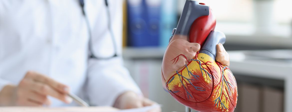
Our cardiology teams provide medical diagnosis and treatment for congenital heart disease, coronary artery disease, heart failure, valvular heart disease and electrophysiology.
CORONARY HEART DISEASE
Our cardiology department is responsible for treating conditions that affect the blood vessels that feed the heart muscle.
Coronary artery disease is a condition in which the coronary arteries become narrowed, reducing or blocking blood flow to certain parts of the heart. This makes it harder for the heart to pump blood to the body.
Coronary artery disease is also known as coronary heart disease or ischaemic heart disease.
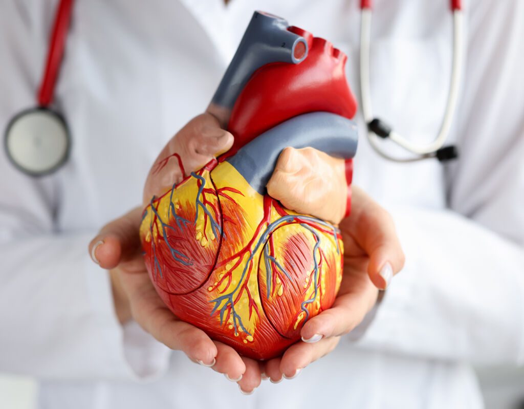

CONGENITAL HEART DEFECTS
This is the treatment of malformations of the heart structure that are present at birth.
Congenital heart defects (treated in our cardiology department) can be mild or severe and can affect one side of the heart or both. The most common type of congenital heart defect is a hole between the chambers of the heart (septal defect).
Other types include holes or defects in the wall between the two main pumping chambers (ventricles) or narrowing of the blood vessels in the heart.
Other less common types include the absence of valves or connections between cavities, abnormal connections between arteries and veins, and abnormal muscle development.
HEART ATTACKS AND STROKES
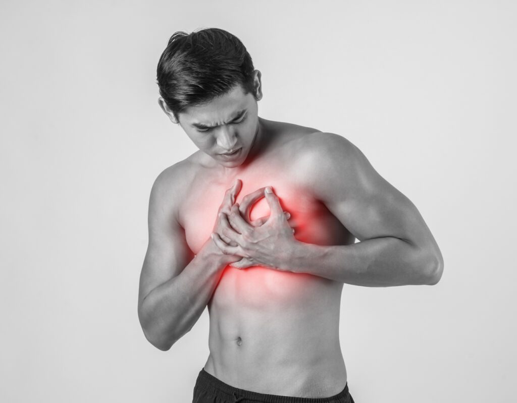
Heart attacks and strokes are usually due to the presence of several associated risk factors such as smoking, poor diet and obesity, physical inactivity and harmful use of alcohol, high blood pressure, diabetes and hyperlipidaemia.
A heart attack is the death of a tissue due to a blockage in the blood supply. A heart attack can be caused by myocardial ischaemia, occlusion of a cerebral artery, kidney damage resembling a heart attack (mossy cell arteriopathy) or other conditions.
Treatment depends on the cause and location of the heart attack. In some cases, surgery may be needed.
CEREBROVASCULAR DISEASE
Treatment of diseases that affect the blood vessels that feed the brain.
These conditions include stroke, brain aneurysm and transient ischaemic attack (TIA).
Cerebrovascular disease (treated in our cardiology department) can be caused by atherosclerosis (hardening of the arteries) or by a tear in the wall of an artery that allows blood to leak into the surrounding brain tissue. This leakage causes a stroke or transient ischaemic attack (TIA).
Atherosclerosis is most commonly associated with high blood cholesterol and smoking. It can also be caused by diabetes and high blood pressure. In some cases, atherosclerosis can occur without any known risk factor.

HIGH BLOOD PRESSURE
Hypertension is a condition characterised by high blood pressure.
Hypertension (high blood pressure) is a long-term condition characterised by an increase in blood pressure. Blood pressure is the force of the blood against the walls of the arteries when the heart pumps blood. Arteries are blood vessels that carry oxygen-rich (red) blood from the heart to other parts of the body.
High blood pressure can lead to serious health problems, including heart disease and stroke. The risk of these problems increases with age and if high blood pressure is not treated over time.
HEART RHYTHM PROBLEMS
Our cardiology department deals with heart rhythm problems. When the heart rate changes for no apparent reason, it is a heart rhythm disorder or it is called an arrhythmia.
Cardiac arrhythmias are part of a group of conditions that involve irregular heartbeats, including arrhythmias, which are disturbances in the frequency or rhythm of the heartbeat.
The most common arrhythmias are atrial fibrillation (AF), atrial flutter and paroxysmal supraventricular tachycardia (PSVT). Although these conditions do not directly cause heart failure, they can increase the risk of other cardiac complications.
WHAT IS A HEART ATTACK?
Myocardial infarction (MI) is a condition treated in our cardiology unit. It is defined as the death of part of the heart muscle due to a lack of blood supply. It is commonly known by its former name “heart attack” and usually has no warning signs. The symptoms of a heart attack can be similar to other medical conditions, making diagnosis difficult.
Myocardial infarction can be caused by coronary thrombosis, a blood clot that blocks a coronary artery, or by a tear in the free wall of the ventricle in the non-criminal cusp. Coronary thrombosis is the most common cause of heart attack. The main risk factor for coronary heart disease is atherosclerosis, which has both genetic and environmental components. Other risk factors include smoking, high blood pressure, diabetes mellitus, lipid abnormalities, obesity and high cholesterol.
The most common symptom of a heart attack is chest pain radiating into one or both arms, with or without shortness of breath, nausea or vomiting. Other symptoms may include dizziness or lightheadedness, palpitations, weakness, tiredness, sweating and a feeling of an extra heartbeat.
WHAT IS CORONARY ARTERY BYPASS GRAFTING?
Coronary artery bypass grafting (CABG) is a surgical procedure that improves blood flow to the heart by diverting blood away from blocked arteries
Coronary artery bypass grafting (CABG) is a surgical procedure that improves blood flow to the heart by diverting blood away from blocked arteries. The surgeon takes blood vessels from other parts of the body and attaches them to the heart and elbows in place. This helps to restore blood flow to the heart muscle, relieve angina and improve heart function.
Coronary artery bypass grafting (CABG) is used to treat coronary artery disease, which occurs when fatty deposits build up in the inner walls of the arteries that supply oxygen-rich blood to the heart.
There are two main types of coronary artery bypass grafting, the most common of which is pump-free coronary artery bypass grafting (PPCAB).
In this type of bypass surgery, surgeons use only their hands during the operation. They do not use mechanical devices such as pumps or stents. This technique has been shown to reduce complications and improve outcomes in patients with stable angina who cannot take medication alone.
Atherosclerosis is a disease of the arteries in which fat builds up and hardens. It is a major cause of coronary heart disease and stroke.
We present to you some articles relating to pathologies treated in our Mediterranean clinic, with an effective and efficient medical and paramedical team, inside our Cardiology Department.
Atherosclerosis is a chronic inflammatory disease of the blood vessels that results from the accumulation of lipids and fibrosis of the arterial wall. This process causes the arterial wall to become thicker, stiffer and harder. The disease affects large and medium-sized arteries throughout the body, particularly those of the heart (coronary arteries), brain (carotid arteries) and legs (peripheral arteries).
The main characteristic of atherosclerosis is the narrowing of an artery due to the build-up of plaque on its inner walls. When this narrowing obstructs blood flow in an artery, it can lead to angina or a heart attack. At a later stage, atherosclerosis can cause peripheral arterial disease, which can lead to gangrene if not treated properly.
Symptoms of atherosclerosis include:
- Tiredness
- Dizziness
- Chest pain
- Leg pain
- Shortness of breath
Lymphoedema (treated in our cardiology department) is a condition that causes swelling of the limbs and other parts of the body. Swelling occurs when lymphatic vessels are damaged or blocked, preventing the body from draining excess fluid.
Lymph vessels are part of the immune system that helps fight infections and diseases. They also collect fluid that returns to the heart, lungs and other organs after passing through tissues such as the skin, nose and mouth. If these vessels are blocked or damaged, they cannot properly drain the fluid they collect into larger veins. This extra fluid accumulates in the affected area and causes swelling.
Swelling caused by lymphatic involvement can be painless or painful, depending on where in the body it occurs. It can also cause itching, redness and heat in the affected area. If you have significant swelling for a long time, it can lead to skin changes such as thickening (papular lymphoedema) or discolouration (late lymphoedema).
There are two types of lymphoedema: primary and secondary.
- Primary lymphoedema occurs when something goes wrong with the lymphatic system itself, such as congenital anomalies (present at birth) or tumours that affect the lymph nodes.
- Secondary lymphoedema occurs when something blocks the flow of lymph fluid through damaged or diseased tissue. This type of lymphoedema most commonly occurs after surgery for breast cancer or some other types of cancer, but it can also occur after surgery to remove an infection from the appendix, gallbladder or tonsils, or after other conditions affecting internal organs.
Treating lymphedema:
If you suspect you have lymphoedema, see your doctor immediately for a diagnosis and treatment options. Lymphoedema should be treated by a doctor who specialises in treating this condition. Your doctor will probably recommend a combination of treatments, including compression bandages and exercises to increase lymph circulation throughout the body.
Compression bandages are usually worn for several weeks until they no longer provide adequate support for your limb(s). At this point, you will be reassessed to determine if additional compression bandages are needed or if custom-made stockings should be used instead.
Dyspnoea is a medical term used to describe shortness of breath. It is an uncomfortable sensation of difficulty breathing that can be associated with physical exertion or even rest.
Dyspnoea can be experienced as chest tightness, difficulty breathing in (inspiratory dyspnoea) or out (expiratory dyspnoea). This is not the same as breathlessness caused by anxiety or panic attacks, which have no physical basis.
Breathing in or out against a closed airway creates resistance to airflow and causes dyspnoea. This occurs in asthmatics who suffer from bronchospasm (narrowing of the airways), but also in patients without respiratory disease who are undergoing surgery in the chest cavity, such as open heart surgery.
Bouveret’s disease is a rare genetic disorder that causes nerve damage in the eye. It is also known as optical atrophy or Progressive external ophthalmoplegia (PEO).
It is caused by a mutation in one of two genes:
BOUVET1, located on chromosome 10q11; and
BOUVET2, located on chromosome 2q33-34.
These genes provide instructions for making proteins called light myosin chains, which are important for muscle function. Mutations affect the ability of light myosin chains to attach to other proteins and form functional muscles. As a result, affected people have reduced muscle strength and weakness in the arms and legs (peripheral neuropathy). They may also have problems with balance and coordination (ataxia), which can affect their ability to walk normally.
In addition to muscle weakness, people with Bouveret’s disease have vision problems that get worse over time. The first symptoms usually appear between the ages of 20 and 40, usually during puberty or early adulthood, but can occur at any age thereafter.
Stenosis is the medical term for the narrowing or constriction of a channel, duct or light. Stenosis can be caused by degenerative diseases such as osteoarthritis and rheumatoid arthritis. In the heart, stenosis refers to a narrowing of the arteries that supply blood to the heart muscle. This can be caused by the build-up of plaque in the arteries (atherosclerosis), which reduces their diameter.
Stenosis can also occur in other vessels leading from the heart, such as the coronary arteries and aorta, and in heart valves. Symptoms include pain, numbness and weakness in the lower back, legs and arms. The pain can be sudden or gradual and can vary in intensity from mild to severe.
Stenosis can be caused by degenerative disc disease or spinal stenosis (also called spinal canal stenosis). Degenerative disc disease occurs when the discs between the vertebrae become thinner and less flexible over time, leading to swelling and herniation of the disc material through the spaces between the bones (vertebral foramina). This herniation puts pressure on the nerves that transmit sensation to the body. Spinal stenosis occurs when the bones of the spine collapse (osteophytes) or move closer together (spondylolisthesis), narrowing the opening of the spinal canal.
Treatment depends on the cause of the stenosis, but may include medications such as steroids or anti-inflammatories, physiotherapy, and surgery to remove damaged bones or clear narrowed spaces in the spine.
Angina pectoris, also known as angina, is a type of chest pain or discomfort that occurs when the heart is not getting enough blood and oxygen. Angina pectoris (treated in our cardiology department) can sometimes be a warning sign of a heart attack.
Angina occurs when the heart muscle needs more oxygen-rich blood than it can get from the coronary arteries. These are the arteries that supply blood to the heart muscle. If they are narrowed or blocked, they cannot supply enough blood to meet the heart muscle’s needs. This causes the symptoms of angina.
There are two main types of angina: stable angina and unstable angina.
Stable angina occurs at rest or during physical activity that does not put significant stress on the heart, and can occur even when the heart is beating normally (normal heart rate). Stable angina may get better with rest, but usually gets worse with physical activity that increases the heart rate or breathing rate (ventilation).
Unstable angina occurs suddenly and severely when the heart needs more oxygen-rich blood than it can get through narrowed or blocked coronary arteries. Unstable angina can occur at rest or during exercise.
In Tunisia, almost 30% of deaths each year are due to cardiovascular disease.
Wondering what the symptoms and risks of high blood pressure are? This guide will provide you with all the information you need to understand and prevent this health problem.
Tachycardia is a heart rhythm disorder that can cause unpleasant symptoms. Learn about the causes, symptoms and treatments in this comprehensive guide.
Pace maker
A pacemaker is a small implanted electronic device that sends electrical impulses to your heart to keep it beating at a normal rate. It detects when your heart is beating too slowly or too quickly and delivers the appropriate electrical impulse to keep your heart beating at a normal rate.
The procedure for implanting a pacemaker varies depending on the location of the heart problem and the type of pacemaker you need. A cardiac electrophysiologist inserts an electrode into the cavities of your heart and connects it to the device. A cable runs from the electrode through the skin and connects to an external power supply carried outside the body.
A pacemaker can be used to treat bradycardia (slow heartbeat) or tachycardia (fast heartbeat).
Bradycardia occurs when the heart beats too slowly, often below 60 beats per minute. It can cause symptoms such as dizziness or tiredness because there is not enough blood circulating around the body. Bradyarrhythmia is rarely life-threatening but can have serious health consequences if left untreated.
Tachycardia occurs when the heart beats too fast, often over 140 beats per minute. It can cause symptoms such as palpitations or shortness of breath because the blood does not have time to fill.
Stent
A stent is a tube placed inside the body to open up an artery and allow blood to flow more easily. It is often used to treat coronary artery disease, which occurs when the heart’s arteries become narrowed or blocked by cholesterol or other substances. Stents come in different shapes and sizes, but they are all made from a strong wire mesh that can be bent into the shape that suits your situation.
If you have coronary artery disease, plaque builds up where two or more arteries meet. This narrowing of the arteries reduces or blocks blood flow to the heart. Stents allow these narrowed areas to be opened up so that blood can flow normally again.
The doctor may recommend stenting if:
- Heart disease caused by blocked arteries (atherosclerosis)
- A very narrow section of an artery (restenosis)
- A blood clot in an artery that hasn’t dissolved on its own.
You ask, our teams answer.
F.A.Q
A brain aneurysm is a weakened and enlarged bulge in a blood vessel. An aneurysm can occur in any artery in the body, but it is most common in the brain.
Brain aneurysms are often called cerebral aneurysms. They are most common in the middle cerebral artery, which supplies blood to the front of the brain, but they can also develop in the arteries that feeding other areas of the brain.
Aneurysms can rupture and bleed into the brain or spinal cord. Blood clots can then form around the aneurysm and block blood flow in the artery. When an aneurysm ruptures, it causes bleeding in or around the brain.
Most people who have a brain aneurysm never have any symptoms. If you experience headaches or other symptoms such as weakness, paralysis, vision problems or changes in personality or behaviour, see your doctor immediately as these could be signs of a potentially serious problem.
Coronary artery disease (treated in our cardiology department) is a condition in which the coronary arteries (arteries that supply blood to the heart muscle) become narrowed or blocked due to the build-up of fatty plaque.
Atherosclerosis is the underlying cause of coronary heart disease. It occurs when plaque builds up in the walls of the arteries. As this plaque builds up, it can shrink or block your arteries. This can lead to angina (chest pain) or a heart attack.
The main symptoms of a heart attack are:
- Chest pain or discomfort
- Pain in other parts of your body, such as your arms, back or neck
- Shortness of breath or trouble breathing
- Nausea and vomiting
- Dizziness or light-headedness.
Atrial fibrillation is a condition in which the upper chambers of the heart (the atria) beat irregularly instead of regularly. This irregular heartbeat can cause blood clots to form in the heart. Clots can break off and travel through the bloodstream to other parts of the body where they can block blood flow, causing tissue damage and possibly death.
Atrial fibrillation can cause blood to accumulate in a part of the heart and prevent it from pumping effectively. This pooling of blood increases the risk of stroke and heart failure.
The most common cause of atrial fibrillation is an irregular heartbeat called an arrhythmia. Arrhythmias are sometimes caused by scarring from previous heart attacks or damage from diabetes. Some people have an inherited tendency to have an arrhythmia. But in many cases, there is no known cause for atrial fibrillation or another type of irregular heartbeat.
It is estimated that around 2% of people aged 65 and over have atrial fibrilation, but it is more common in men than women and most common after the age of 65.
A transient ischaemic stroke, also known as a mini-stroke, is a temporary interruption in the blood supply to the brain. It can be caused by a blood clot or a ruptured blood vessel.
Transient ischaemic events are often called mini-strokes because they have some of the symptoms of a full stroke. But they are usually shorter and less severe than a classic stroke.
A transient ischaemic attack (TIA) is an episode that lasts less than 24 hours and causes sudden symptoms such as:
- Weakness or numbness in the face, arm or leg, especially on one side of the body
- Difficulty speaking
- Confusion or difficulty understanding what people are saying
- Loss of vision in one or both eyes.
- Dizziness or loss of balance.
Ventricular septal defects (take in charge within Cardiology department)are a group of congenital heart defects that affect the separation between the left and right sides of the heart. Some babies with these defects have no signs or symptoms before birth. Others have signs or symptoms, such as an abnormal heart rhythm (arrhythmia) or shortness of breath.
There are different types of septal defect. Your doctor can tell you which type you have. The most common types are:
Interventricular communication (IVC): Interventricular communication is a hole in the muscle wall between the ventricles (lower chambers) of your heart. This allows oxygen-rich blood to pass from one ventricle to another instead of going to the lungs, reducing the amount of blood circulating there. IVCs range from small holes that can close on their own to larger holes that require surgery.
Interauricular communication (ICD): An ICD is a hole in the wall between the upper chambers (atria) of your heart. It allows oxygen-rich blood to pass from one atrium to the other instead of going to the lungs.
Pericarditis (take in charge within Cardiology department) is an inflammation of the pericardium (a condition treated in our cardiology department). It can be acute and self-limiting, or it can become chronic.
The pericardium is a two-layered bag that surrounds the heart, providing protection and allowing blood to flow from the heart to the body. Pericarditis occurs when part of the pericardium becomes inflamed. Pericarditis can occur as a result of infection (viral, bacterial or fungal), inflammation associated with cancer treatment, autoimmune disease, radiotherapy for cancer and chest trauma.
Symptoms of pericarditis:
Symptoms of acute pericarditis include:
- Chest pain (sharp and throbbing) in the centre or near the sternum (breastbone) that gets worse with deep breathing or movement.
- Pain that spreads to the shoulders, arms and back (flank)
- Persistent fever
- Chills
- Tiredness
- Swelling of the neck (especially on one side) if you have associated symptoms of pleurisy.


