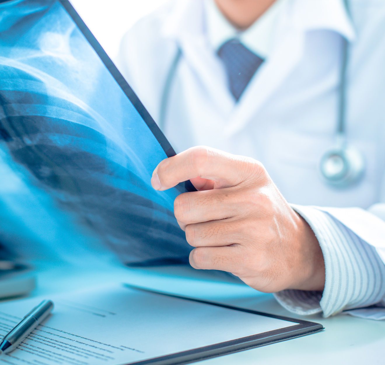
Radiology, or radiological examination, is the process of using ionising radiation and its interaction with matter to produce images of the inside of the body.
In general, radiological examinations are used to facilitate diagnosis and may also be used to guide treatment. Radiological interventions, such as therapeutic procedures, can be guided by the anatomical information provided by imaging techniques.
Radiological examinations include x-rays, CT scans, fluoroscopy, magnetic resonance imaging (MRI), ultrasound and nuclear medicine imaging techniques.
The word ‘radiology’ sometimes refers to medical imaging in general, sometimes to the image acquisition aspect of medical imaging, sometimes to nuclear medicine and other non-radiological diagnostic procedures but is often considered synonymous with medical imaging in general.
Radiological examination (radiology) is an important diagnostic procedure in medicine and dentistry.
All the teams in our clinic are committed to ensuring the quality and safety of care on a daily basis.
120 Hospital beds, 32 Outpatient beds, 15 Operating theaters, 128-strip scanner, Ultrasound, Doppler, …
Emergency service, reception 24 hours a day, 7 days a week, with personalized care for all our patients.
Our radiology department uses a 128-slice scanner. It is one of the most important diagnostic tools used in medical practice. It looks for abnormalities that are not visible to the naked eye, including tumours and internal bleeding.
- Ultrasound is a method of creating images using sound waves in the form of ultrasonic pulses. It has many uses in modern medicine and other applications but is perhaps best known for its use in obstetric diagnosis.
Ultrasound has many applications in medicine and dentistry. It is often used to examine: - The abdomen (the belly)
- The heart, including valves and cavities
- Blood vessels and circulation in the heart and brain (cardiac imaging)
- The liver, gallbladder, spleen, kidneys and urinary tract (abdominal ultrasound)
- The pelvis (pelvic ultrasound)
The Doppler method uses the frequency shift caused by the Doppler effects to measure blood flow in arteries and veins. Typically, this method involves using a probe to send high-frequency sound waves into the artery or vein, and then measuring their frequency as they bounce back. The probe records these frequencies at several points in the vessel and then uses standard formulae to calculate changes in blood velocity and volume.
Conventional radiology is a branch of medicine that uses imaging techniques to facilitate the diagnosis, monitoring and treatment of medical conditions. Specific examples of the techniques used are radiography (X-ray), computed tomography (CT), magnetic resonance imaging (MRI) and ultrasound.
Radiography is a diagnostic test that uses X-rays to visualise internal structures.
X-rays are a form of electromagnetic radiation (EMR). Electromagnetic radiation is energy that travels in waves and can only be seen by the human eye. X-rays have much higher energy than visible light, but lower energy than ultraviolet rays and other types of EMR such as gamma rays, radio waves and microwaves.
X-ray machines use high voltage to produce X-rays that pass through the body. When X-rays pass through a material, they create shadows on a photographic film or digital detector called an image intensifier. The denser the material, the stronger the shadow on the film or intensifier screen.
Diagnostic medical X-rays are taken for many reasons; including:
- Seeing fractures or breaks in bones;
- Diagnose tumours;
- Check for foreign bodies in the body;
- Assess injuries that require surgery
Magnetic resonance imaging (MRI) is a medical imaging technique used to visualise the internal structures of the body. MRI uses magnetic fields and radio waves to produce detailed images of the body’s organs and other internal structures.
An MRI can be used to diagnose a number of conditions, including:
- Problems with the brain and spinal cord, such as tumours, cysts and arteriovenous malformations (AVMs).
- Tumours of the spine or brain, including meningiomas, pituitary adenomas and acoustic neurinomas.
- Spinal problems, such as herniated discs or spinal stenosis (narrowing of the spinal canal)
- Sciatica or other nerve problems
A specialised radiological examination (part of the radiology department) is an examination that requires special training or equipment. Diagnostic radiologists have undergone years of education and clinical training to understand the biology of diseases and how they appear on imaging tests, and to correctly interpret and apply the information they see on a particular test.
OUR SERVICES
Radiological examinations in our radiology department include x-rays, CT scans, fluoroscopy, magnetic resonance imaging (MRI) and ultrasound. Here are some more explanations:
MRI
Magnetic resonance imaging (MRI) is a non-invasive medical test that uses magnetic fields and radio waves to create images of your body. MRI is often used to diagnose diseases such as cancer and structural problems in the bones, joints, spine and brain.
Scanner
Medical scanners are used in many types of medical procedures. They provide images of internal organs and other parts of the body to determine if there are problems or abnormalities. Medical scanners are also known as imaging equipment or radiology equipment. The type of equipment used depends on the specific procedure being performed and the type of information needed from the scanner.
X-RAYS
X-rays are a form of electromagnetic radiation (a type of energy) similar to light, but with a shorter wavelength. X-rays can be produced artificially using X-ray tubes, which are devices that produce high-intensity X-rays by accelerating electrons to high speeds in a vacuum. X-ray machines are used in hospitals and other medical facilities to take pictures of the inside of the human body. These images are used for diagnostic purposes, particularly to detect fractures or internal bleeding.
MRI
MRI (Magnetic Resonance Imaging) is a medical imaging technique that uses a powerful magnet, radio waves and a computer to produce detailed images of the body.
MRI scanners have become commonplace in hospitals and medical clinics around the world. MRIs are used to diagnose many types of illness and injury, including cancer, heart disease, stroke, brain injury and bone disease.
MRI can also be used to monitor the development of an injury or disease over time. This allows doctors to follow their patients more closely and ensure that their condition is improving or deteriorating at the right rate.
MAMMOGRAPHY
Mammography is a medical imaging technique used to help diagnose breast cancer and other breast diseases. During a mammogram, an X-ray machine sends a beam of radiation into the breast. The X-rays pass through the breast and expose the film. Your doctor will use the images to look for signs of cancer or other conditions.
Mammograms are also used to check the effectiveness of treatments. For example, it can be used to check whether tumours have changed in size or shape after drug treatment or radiotherapy.
ANGIOGRAPHY
Angiography is a medical imaging technique used to visualise and provide information about the body’s vascular system. It involves injecting a radioactive contrast agent, usually a linear polymer solution, into the bloodstream and x-raying it as it travels through the blood vessels. The procedure produces images of the blood vessels, allowing doctors to detect abnormalities in the structure or flow of the vessels.
Angiography can be used to detect certain types of cancerous tumours in the liver, pancreas, adrenal glands and lungs. It can also be used to detect heart disease and coronary artery disease (atherosclerosis).
OUR TEAM WILL ANSWER YOUR QUESTIONS
F.A.Q
A scan of the stomach is needed to confirm the diagnosis and improve the prognosis. It is done with a contrast medium. X-rays must be taken before any surgical procedure.
An X-ray of the stomach can show:
– The size and shape of the organ;
– the various changes in its shape;
– the presence or absence of ulcers, tumours and other pathological
– Lesions;
– perforations;
– Obstruction of blood vessels (angiodysplasia).
Radiological examination of the stomach is a common procedure. It can be used to detect the presence of tumours, bleeding or other abnormalities.
The most common radiological test is a barium swallow. This involves swallowing a thick liquid called barium sulphate, which coats the inside of the mouth and throat. X-rays are taken to look at the digestive tract. Barium swallowing can also be used as a precursor to an endoscopy.
Radiological tests of the kidneys and urinary tract in children include ultrasound, intravenous urography (IVU) and computed tomography (CT).
Ultrasound is used to look for abnormalities in the kidneys and urinary tract. It is performed using a special examination technique that gives a clear picture of these organs. Ultrasound can be used to:
• Detect malformations of the urinary tract;
• Assess micturition problems;
• Detect tumours at an early stage.
Kidney and urinary tract infections in children are often associated with pain, fever, nausea and vomiting. The condition can be acute or chronic. Acute pyelonephritis is characterised by a sudden onset of symptoms and often requires urgent hospitalisation.
The diagnosis of pyelonephritis is based on the results of a urinalysis, which shows an increase in white blood cells and bacteria in the urine. In addition to inflammation of the kidneys, the disease may be associated with inflammation of other organs (renal abscesses).
Portal hypertension is a condition in which the blood pressure in the portal vein is higher than normal. It occurs when there is an increase in pressure in the portal vein due to a blockage in its flow. The portal vein carries blood from the gastrointestinal tract, spleen and pancreas to the liver.
Radiological investigation of portal hypertension
Portal hypertension can be diagnosed using simple imaging techniques such as ultrasound and CT scans. The results can then be correlated with other clinical signs such as abdominal pain, jaundice and weight loss.
Right and left upper quadrant ultrasound (RUQUS) is one of the most common radiological tests used to evaluate patients with suspected or proven portal hypertension. This is a non-invasive technique for assessing the size and shape of the liver and spleen. The usefulness of RUQUS lies in its ability to detect abnormalities such as splenomegaly, ascites and free fluid around the abdominal cavity. It can also be used to assess the vascularity of these organs.
Nos autres centres d'explorations

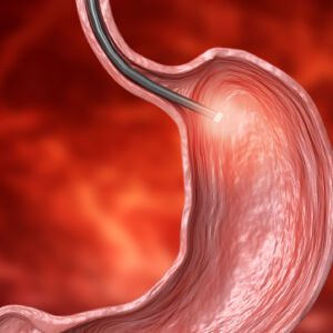
Gastroscopy is an invasive medical procedure used to diagnose and treat diseases of the gastrointestinal tract.
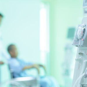
Dialysis is a procedure in which a machine is used to replace kidneys that have stopped working. Dialysis can be used by patients with kidney failure, kidney disease or perioperative surgery.

In medicine, cardiac exploration is a surgical procedure involving the heart.

Biological testing is a general term that covers many different types of tests that can be performed on blood, urine and tissue samples.