In medicine, cardiac exploration is a surgical procedure involving the heart.
Cardiac explorations are frequently performed on patients of all ages to repair damaged tissue around the heart and improve their overall quality of life.
The definition of cardiac exploration is a procedure that involves the coronary arteries and other blood vessels connected to the heart.
To better define cardiac exploration, it needs to be broken down into its different parts. The first part of cardiac exploration is coronary angiography, where surgeons work on the coronary arteries to correct damage or blockages caused by atherosclerosis.
The second part of the heart scan is angiography, where a catheter is used to access the blood vessels leading to and from the heart.
The final part of the cardiac examination is pericardiocentesis, where surgeons work on the pericardium – the sac that surrounds the heart – to repair any tears or tears that may be present.
All the teams in our clinic are committed to ensuring the quality and safety of care on a daily basis.
120 Hospital beds, 32 Outpatient beds, 15 Operating theaters, 128-strip scanner, Ultrasound, Doppler, …
Emergency service, reception 24 hours a day, 7 days a week, with personalized care for all our patients.
Rhythmic Holter is a complete examination of the electrocardiogram and automatic analysis to detect changes in the heart rate. It is performed over 24 hours to study the heart rate during all daily activities.
Arrhythmias are abnormal rhythms that can cause the heart to beat faster or slower than expected. A person with an arrhythmia may feel dizzy, weak or have palpitations (a feeling of the heart beating). Arrhythmias can disrupt blood flow to the heart and brain and cause serious complications if left untreated.
Cardiac arrest occurs when the heart’s electrical activity suddenly stops and its pumping action becomes ineffective – sometimes within seconds. Cardiac arrest is a life-threatening emergency because it deprives the brain and other vital organs of the oxygen-rich blood they need to function properly.
Holter tensionnel is a French term that literally means “tension du jour”. In order to determine whether or not a patient has a true arrhythmia, it is necessary to rule out the possibility that the symptoms are caused by stress. The Holter blood pressure test is a stress test.
The Holter monitor is a portable recording device, usually worn on a belt or shoulder strap that continuously records electrocardiogram (ECG) waveforms and events for up to 24 hours. The Holter monitor can be programmed to record all or part of the five different leads, as well as rhythm bands, arrhythmia detection events, blood pressure measurements and voice recordings of the patient’s symptoms.
Exercise testing is a procedure used to determine whether a person has exercise-induced cardiomyopathy or an ICD. It involves monitoring the electrical activity of the heart while the patient is exercising on a treadmill, stationary bicycle or other equipment.
The results of a stress test show whether your heart gets enough blood flow during exercise and whether there are areas of the heart muscle where blood flow is inadequate. If the arteries in the heart muscle are blocked, blood flow will be lower than normal when you exercise or experience extreme emotional or physical stress. This can cause symptoms such as chest pain, shortness of breath and tiredness.
Cardiac ultrasound is a medical imaging technique that uses high-frequency sound waves to create an image of the heart and surrounding structures. The resulting image (or ‘echocardiogram’) can be used to measure the size and other characteristics of the heart’s chambers, valves and walls. It can also show leaks in the heart, damage or abnormalities in the valves or walls, or the movement of blood in an area outside its normal path.
Cardiac ultrasound can help your doctor diagnose and treat many heart conditions, including:
– Arrhythmias (irregular heartbeats): Arrhythmias are abnormal heart rhythms. The most common symptoms are palpitations, shortness of breath, dizziness and fainting.
– Heart valve problems: Heart valve problems are conditions that affect the heart valves. Valves are pieces of tissue that open and close to allow blood to flow in one direction through the heart chambers and vessels. Valvulo diseases can be genetic or acquired.
– Congenital heart disease (problem at birth): Congenital heart disease is a term used to describe a wide range of defects in the structure of the heart. Congenital heart defects are present at birth, but are not always obvious. Some types of congenital heart disease are diagnosed shortly after birth, while others are not diagnosed until adulthood.
– Heart muscle problems (cardiomyopathy):
Cardiomyopathy is a disease of the heart muscle that can lead to abnormal heart function. The heart muscle, also called myocardium, is made up of a strong outer layer and a soft, blood vessel-filled centre. In cardiomyopathy, the myocardium becomes thicker and wider, making it harder to pump blood through the heart.

WHAT IS AN ELECTROPHYSIOLOGICAL STUDY?
Electrophysiological testing is a medical treatment used for patients with arrhythmias.
Electrophysiological testing uses an electrical stimulus to create a potential for action in heart tissue. The electrophysiologist records the resulting electrocardiogram (ECG) and sometimes other signals.
The procedure is also called an EP study, an electrophysiological study (EPS) or sometimes just “EP”.
The procedure:
An electrophysiological study is the medical treatment of an arrhythmia using a device that delivers electrical impulses to the heart. The term can refer to any of the following:
A procedure in which a catheter is placed in the heart and used to record the electrical activity of the heart muscle. A device attached to the catheter that records and displays the activity on an electrocardiographic (ECG) monitor.
A procedure in which a catheter is inserted into the blood vessels to deliver medication directly to the heart muscle. This procedure may be performed instead of or in addition to open-heart surgery.
OUR TEAM WILL ANSWER YOUR QUESTIONS
F.A.Q
Coronary angiography is a medical procedure that uses X-ray imaging to examine the heart and coronary arteries. Coronary arteries are the blood vessels that supply oxygen-rich blood to the heart muscle.
Coronary angiography can help doctors diagnose and treat a number of heart conditions:
- Heart attack: If you have chest pain, shortness of breath or other symptoms that may indicate a heart attack, coronary angiography can help determine if your pain is caused by coronary artery disease. This condition occurs when fatty deposits build up in the walls of the coronary arteries, blocking blood flow.
- Angina pectoris (chest pain). Angina pectoris occurs when an insufficient amount of oxygen-rich blood reaches the heart muscle due to coronary artery disease or other causes. Coronary angiography can be used to rule out other causes of angina pectoris as well as to diagnose and treat angina.
Angiography is an invasive, X-ray-based medical procedure that uses a catheter to take pictures of the blood vessels. The catheter is inserted into the blood vessel and advanced until its tip reaches the area of interest.
A contrast dye is injected into the bloodstream through the catheter and images are taken as the dye moves through the blood vessels.
Angioplasty may be carried out during angiography. This involves inserting a balloon into an artery blocked by atherosclerosis (plaque) and inflating it to open the blocked artery.
Nos autres centres d'explorations


Radiology, or radiological examination, is the process of using ionising radiation and its interaction with matter to produce images of the inside of the body.
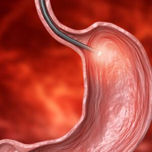
Gastroscopy is an invasive medical procedure used to diagnose and treat diseases of the gastrointestinal tract.

Dialysis is a procedure in which a machine is used to replace kidneys that have stopped working. Dialysis can be used by patients with kidney failure, kidney disease or perioperative surgery.

Biological testing is a general term that covers many different types of tests that can be performed on blood, urine and tissue samples.